
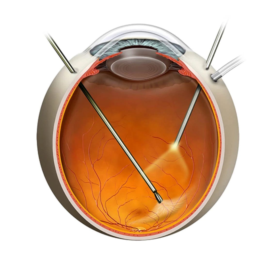
Vitrectomy is a modern microsurgical procedure used to treat conditions affecting the vitreous and the retina. During the procedure, the vitreous body—the transparent, gel-like substance that fills the inside of the eye—is removed using very fine microsurgical instruments. This allows the surgeon direct access to the retina and enables precise treatment of the underlying condition.
The procedure is usually performed under local anaesthesia, and in most cases, the patient can return home on the same day. Depending on the underlying condition, air, a special gas or silicone oil may be placed inside the eye to support the retina during the healing process. Following surgery, temporary blurred vision, redness or mild discomfort may occur, while in some cases, a specific head position may be required for several days. The duration of recovery and the degree of visual improvement depend primarily on the underlying condition and the initial state of the retina.
Who it is for
Vitrectomy is used to treat conditions affecting the vitreous and retina when surgical intervention is required to preserve or improve the anatomy and function of the eye. It is used, among other indications, in cases of retinal detachment, vitreous haemorrhage, epiretinal membrane, macular hole, vitreoretinal traction and complications of diabetes. The need for vitrectomy is assessed on an individual basis, depending on the underlying condition and the specific characteristics of each eye.