
Dr. Asterios Diafas
Ocular OncologyVitreoretinal Surgery
+30 694 240 6078asterisdiafas@hotmail.com
Condition · Ocular oncology

Choroidal hemangioma

A benign vascular lesion of the choroid

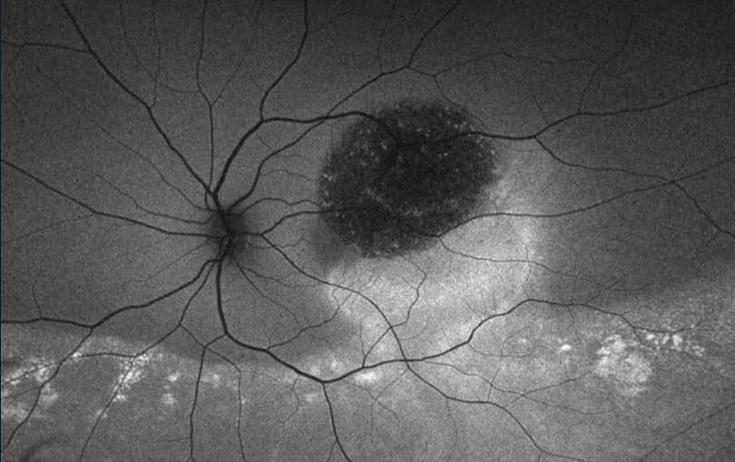
Clinical image — Choroidal hemangioma.
Overview

A choroidal hemangioma is a benign vascular tumour of the choroid, the highly vascular layer located beneath the retina. It usually develops slowly and may cause no symptoms.

When located near the macula or when it causes fluid to accumulate beneath the retina, it can affect vision. Most choroidal hemangiomas are solitary and occur in one eye.

Symptoms

Many patients have no symptoms, and the lesion is discovered incidentally during a routine eye examination. When it affects vision, symptoms may include:

  • Blurred or reduced central vision
  • Distortion of straight lines
  • A dark or blurred area in the visual field
  • Reduced colour perception

Symptoms are usually related to the presence of fluid beneath the retina.

Risk factors

The exact cause of choroidal haemangioma is not fully understood. Most cases occur sporadically, without a clearly identifiable underlying cause. They have been associated with congenital vascular abnormalities and, particularly in diffuse forms, with Sturge-Weber syndrome. Choroidal haemangiomas may be present from birth, while in many cases no specific risk factor or underlying condition can be identified.

How is it diagnosed?

The diagnosis is based on a comprehensive eye examination and specialised imaging tests. OCT (Optical Coherence Tomography) helps assess the retina and detect any associated fluid.

Ocular ultrasound and angiography can help confirm the diagnosis and distinguish a choroidal hemangioma from other choroidal lesions.

Treatment

If a choroidal hemangioma does not cause symptoms or complications, regular monitoring may be all that is required.

When it causes vision loss or fluid accumulation beneath the retina, treatment may be necessary. Photodynamic therapy (PDT) is one of the main treatment options and can help reduce the lesion and associated fluid, with the aim of improving or preserving vision.

When to seek an assessment

Schedule an eye examination if you notice a new or gradual change in your central vision, distortion of straight lines, or a blurred area in your visual field. If your ophthalmologist identifies an intraocular lesion, you may be referred to a specialist ophthalmologist with expertise in ocular oncology for further assessment.

Sudden significant loss of vision, the new onset of flashes or floaters, or the appearance of a dark “curtain” in your visual field requires urgent ophthalmic assessment, as these symptoms may be associated with another eye condition requiring immediate attention.

Key points
  • It is a benign vascular tumour and often causes no symptoms.
  • Blurred or distorted central vision may occur.
  • Modern imaging techniques improve the diagnostic accuracy and reliable monitoring of the lesion.
  • Photodynamic therapy (PDT) is an important treatment option.

Talk it through with Dr. Diafas.

English: For an evaluation, careful monitoring, or a second opinion, book your appointment.

This page provides general information only and is not a substitute for an individual consultation.

Book an appointment