Dr. Asterios Diafas
Ocular OncologyVitreoretinal Surgery
+30 694 240 6078asterisdiafas@hotmail.com
Condition · Medical retina

Retinal vein occlusion

A vascular occlusion affecting vision

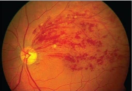
Clinical image — Retinal vein occlusion.
Overview

Retinal vein occlusion is a vascular condition in which the normal flow of blood through a retinal vein is obstructed. The occlusion can cause retinal haemorrhages, macular oedema, and visual disturbances.

Depending on the vessel affected, it is classified as central retinal vein occlusion (CRVO) or branch retinal vein occlusion (BRVO).

Symptoms

Symptoms may include:

  • Sudden or gradual blurred vision
  • Reduced central vision
  • Distortion of vision
  • A visual field defect
  • Sometimes no symptoms, particularly with a small branch vein occlusion

RVO is usually painless.

Risk factors

Retinal vein occlusion is commonly associated with vascular risk factors such as hypertension, diabetes mellitus, elevated cholesterol, smoking, and older age, particularly in patients with cardiovascular disease or other cardiovascular risk factors. In younger patients, however, the underlying causes may differ and can include thrombophilic disorders, antiphospholipid syndrome, hyperhomocysteinaemia, autoimmune diseases (such as systemic lupus erythematosus and Behçet’s disease), inflammatory vasculitis, myeloproliferative disorders, use of hormonal contraceptives, glaucoma, or pregnancy. Identifying and managing these risk factors is important for both ocular and overall health. In younger patients, further haematological or rheumatological evaluation may be appropriate depending on the clinical history and findings.

How is it diagnosed?

Diagnosis is based on a detailed ophthalmic examination and retinal imaging. Fundus photography helps document retinal haemorrhages and vascular changes, while macular OCT allows the detection and monitoring of macular oedema.

In selected cases, OCT angiography or fluorescein angiography may be required, particularly to assess retinal blood flow and ischemia.

Treatment

Treatment depends on the type and severity of the occlusion, as well as the presence of complications. When macular oedema is present, the main treatment option is usually intravitreal anti-VEGF injections, which can reduce the oedema and help preserve or improve vision.

In some cases, intravitreal corticosteroids may also be used. If abnormal new blood vessels (neovascularisation) develop, laser photocoagulation may be required, depending on the findings.

At the same time, it is important to identify and manage any underlying vascular risk factors.

When to seek an assessment

If you experience new or sudden blurred or reduced vision, it is important to have an ophthalmic examination. Early diagnosis allows assessment of the retina and macula and helps determine the most appropriate treatment.

If you have been diagnosed with retinal vein occlusion, regular monitoring with OCT is important for the early detection of macular oedema or other complications.

Key points
  • It is a vascular condition that obstructs the normal flow of blood through one of the retinal veins and usually presents with painless reduction in vision.
  • It can cause retinal haemorrhages and macular oedema.
  • OCT helps with the diagnosis, assessment, and monitoring of the condition.
  • Intravitreal anti-VEGF injections, as well as other treatment options, can, in appropriate cases, reduce macular oedema and contribute to improving or stabilising vision.

Talk it through with Dr. Diafas.

For assessment of a sudden change in vision, monitoring of the occlusion, or discussion of treatment with intravitreal injections and laser, book an appointment.

This page provides general information only and is not a substitute for an individual consultation.

Book an appointment